
Introducing: Tip of the Month
Introducing our newest initiative – the ‘Tip of the Month’ feature on the Northwest Region Prehospital Care Program’s Learning Management System. Activate the QR code below for monthly insights, valuable tips, and expert guidance to enhance your prehospital care skills. Elevate your learning experience with our curated tips designed to empower and inform. Let the journey to continuous improvement begin!
Tip of the month
July 2026 - Tip of the Month - Early Transport Decisions in Cardiac Arrest
Early Transport Decisions in Cardiac Arrest
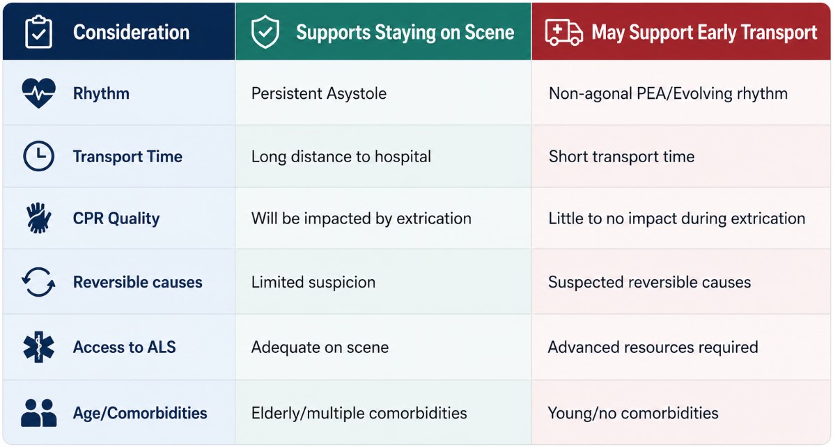
Out-of-hospital cardiac arrest management requires paramedics to balance the benefits of on-scene resuscitation with the potential advantages of rapid transport. High-quality CPR, rhythm analysis, defibrillation, and airway management are often best performed in a controlled on-scene environment. However, some patients who are unlikely to meet Termination of Resuscitation (TOR) criteria may benefit from early transport, and this is not limited to the H’s and T’s of reversible causes. The decision to transport prior to 20 minutes of resuscitation should be guided by objective clinical factors and the ability to maintain high-quality resuscitation throughout extrication and transport.
By the end of this tip, paramedics will be able to identify key clinical factors that should guide decisions regarding early transport during cardiac arrest resuscitation.
You are managing a 54-year-old patient in cardiac arrest with persistent, narrow complex PEA. High-quality CPR is ongoing with an automated CPR device, the airway is managed with a supraglottic airway, there are ample resources on scene for a simple extrication and transport time to the nearest emergency department is less than five minutes. Do you remain on scene to continue resuscitation for 20 minutes, or initiate early transport?
Deciding to leave the scene prior to 20 minutes of resuscitation should never be based on intuition, pressure from bystanders, or a desire to “do more.” Decisions should be based on objective clinical findings and what is most beneficial for the patient.
Consider the full clinical picture. Factors that may influence the decision to transport early include: Suspected cause of arrest, patient age, distance to hospital, access to ACLS interventions, presenting and evolving cardiac rhythm, reversible causes, and ability to maintain high quality CPR during movement and transport.
High quality CPR remains the priority. Transporting a patient should not come at the expense of effective resuscitation. If CPR quality, rhythm interpretations, ventilation or medication administration will be significantly compromised during extrication or transport, remaining on scene may provide the patient with the best opportunity for survival.
Paramedics may initiate transport before 20 minutes of on-scene resuscitation when the decision is supported by objective clinical factors and high-quality resuscitation can be maintained during extrication and transport. The decision must not be based solely on intuition, time pressure, or external pressure.
During your next cardiac arrest call, ask yourself: “will transporting this patient improve access to meaningful interventions without compromising resuscitation efforts?”
June 2026 - Tip of the Month - Stroke Care Excellence
Mastering the Clock and the Card: Optimizing Field Stroke Assessments & Bypass Protocols
When an acute ischemic stroke occurs, millions of neurons die every minute, making rapid reperfusion therapy the highest priority. The Paramedic Prompt Card for Acute Stroke Bypass Protocol (see image below) acts as the guideline for this life-altering care, assisting paramedics to determine when to bypass local hospitals in favor of designated Regional Stroke Centers. However, the success of this hinges entirely on the quality of field assessment by paramedics. Accurate Identification of the Last Seen Normal (LSN) window, swift calculation of large vessel occlusion (LVO), and medical history are what allow the emergency department stroke teams to immediately assess and determine the appropriate treatment including the administration of thrombolytic medication like Tenecteplase (TNK) or other potential treatment pathways.
By the end of this tip, paramedics will be able to accurately calculate and document a patient’s LSN time, correctly apply the LAMS score for LVO screening, and gather the critical anticoagulant data required to optimize the acute stroke bypass.
May 2026 - Tip of the Month - Amputations
Amputations are high-acuity, time-sensitive injuries that can occur in a wide range of environments; from industrial and farm machinery to motor vehicle collisions, power tools, and even sharp-force trauma. The mechanism can vary significantly, from clean surgical-like separations to severe crush or avulsion injuries, each presenting unique challenges for prehospital care. Regardless of the cause, early recognition and proper management are critical to patient outcomes. Effective hemorrhage control, preservation of the amputated part, when possible, and rapid transport decisions all play a vital role in improving survival and the potential for reattachment.
This month’s Tip of the Month is intended to support paramedics in assessing and prioritizing management of traumatic amputations, including effective hemorrhage control and appropriate preservation of the amputated part, in accordance with Ontario BLS and ALS-PCS standards.
Tip of the Month - April 2026 - Glasgow Coma Scale
"Can you open your eyes for me?"
If a patient opens their eyes when you speak to them, are they automatically a GCS 14?
Not necessarily. Accurate scoring depends on how and why the response occurs